Research
Research
Research Interests
The primary research focus of the Waterhouse laboratory is to understand the role of the central monoaminergic systems in brain function and behavior. More specifically, the lab is concerned with the anatomy and physiology of the brainstem noradrenergic and serotonergic efferent systems as they relate to executive function and the sensory-processing capabilities of an organism.
These studies employ a broad spectrum of behavioral, neuroanatomical, electrophysiological, and molecular techniques including microiontophoresis; single and multi-unit extracellular recording from anesthetized animals; simultaneous spike train recordings from multiple arrays of single neurons in waking animals; computer-based acquisition and analysis of spike train data; behavioral paradigms for signal detection, sustained and flexible attention; mapping of monoamine projections from source nuclei using retrograde tracer substances, immunohistochemistry, intersectional genetic models, and viral vector strategies; molecular phenotyping of laser captured monoaminergic neurons using qrtPCR.
The underlying theme of this work is that synaptically released norepinephrine and serotonin operate as complementary neuromodulatory substances that regulate the responsiveness of sensory neurons, sensory circuits, and sensory networks to synaptic inputs. As such, these systems may play a significant role in the ability of the organism to orient and attend to novel or salient stimuli from the sensory surround. More recent work focuses on monoaminergic regulation of prefrontal cortical circuit physiology and executive function - planning, attending, deciding.
Clinical implications of this work which have led to related experimental studies are that these monoaminergic systems may underlie some of the behavioral actions of psychostimulant drugs such as cocaine and methylphenidate (Ritalin ®) and the cognitive deficits that accompany normal aging, anxiety/PTSD, HIV neuroAIDS, traumatic brain injury and attention disorders such as ADHD.

Locus Coeruleus - Norepinephrine System
The laboratory has a longstanding interest in how the norepinephrine released from neurons in the locus coeuruleus influences sensory and cognitive function.
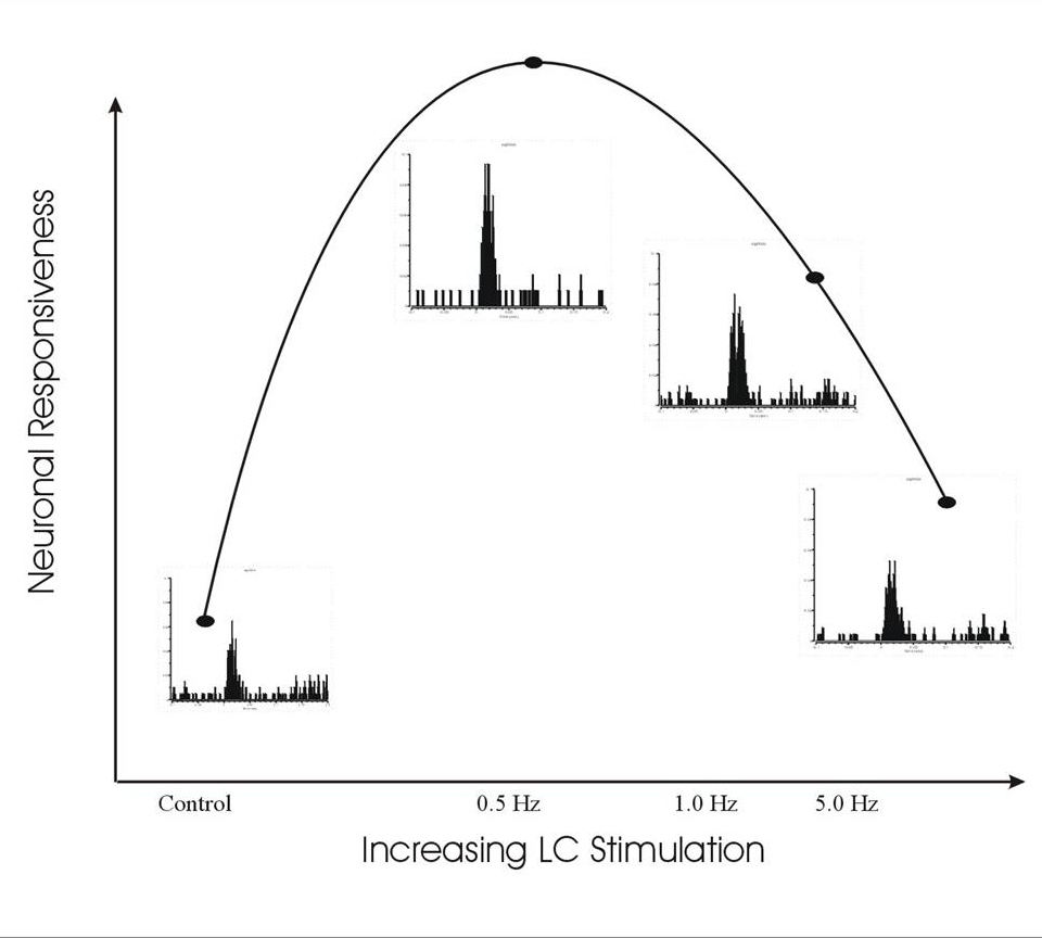
In this figure, electrical stimulation of the locus coeruleus-norepinephrine efferent pathway at different frequencies modulates the sensitivity of a single neuron in the somatosensory thalamus to whisker stimulation, according to an "inverted-U" stimulus-response relationship.

Dorsal Raphe
The brainstem dorsal raphe nucleus (DRN) utilizes multiple neurotransmitter systems in mediating a variety of adaptive and maladaptive behaviors including stress, anxiety, depression, sleep, feeding, pain and nociception.
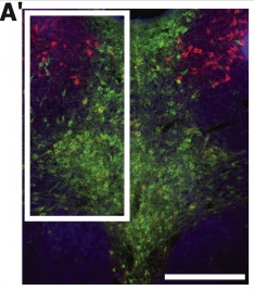
In this horizontal section from a rat brainstem, neurons that synthesize and release serotonin (green) are concentrated along the midline of the raphe, while neurons that synthesize the putative transmitter, nitric oxide (red) are concentrated in the adjacent lateral dorsal tegmental nucleus.

Psychostimulant Drug Actions
Psychostimulant drugs such as methylphenidate (Ritalin ®) are used to treat ADHD but have also become popular for their performance enhancing effects in otherwise normal individuals.
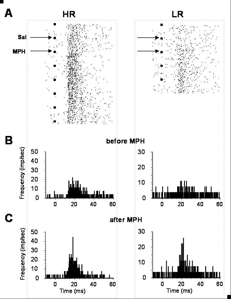
In this figure, two sensory thalamic neurons (HR and LR) from normal rats show more robust responses to sensory-driven synaptic inputs after MPH administration (A,B,C). HR = initially high responding unit, LR = initially low responding unit; Sal = saline, MPH = methylphenidate at 2 mg/kg I.P.

Traumatic Brain Injury
Under normal conditions the norepinephrine (NE) transmitter system regulates flexible attention, a cognitive function that is critical for management of everyday tasks but also highly susceptible to head injuries. Ongoing studies are focusing on the effects of repeated concussive events (mild traumatic brain injury - TBI) on flexible attention and the functionality of the NE transmitter system in the brain. This investigation relies on a rat model of mild TBI and established behavioral, anatomical and electrophysiological assays to link concussion-induced deficits in flexible attention to injury-related pathologies of the NE system.
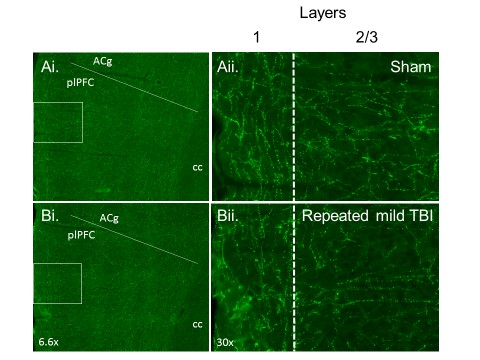
The figure illustrates the density of NE fiber staining in the layers 2/3 of the prefrontal cortex following experimentally-induced repetitive mild TBI. Low- (left) and high- (right) power images show NE fiber staining in the pre-limbic prefrontal cortex of sham (Ai-ii) and repetitive mild TBI (Bi-ii) animals.
