Physiology Project
Physiology Project
Physiology Project
While our work and the work of others have made significant progress on understanding how VSP functions on a biophysical level, much is still unknown regarding the physiological role of VSP. Many phosphatases were already known that can dephosphorylate either the 3- or the 5-phosphates from PIPs. Why does the cell require another one that is regulated by voltage?
To answer this question, we investigated native expression patterns of VSP. We found VSP in the brains and kidneys of frogs among other areas. Looking more closely at the kidney, we discovered that VSP expresses on the apical membrane of the proximal tubule kidney cells (Figure 1, 2). This location suggests VSP could play a role in the ability of these cells to reabsorb nutrients, a critical function of kidneys.
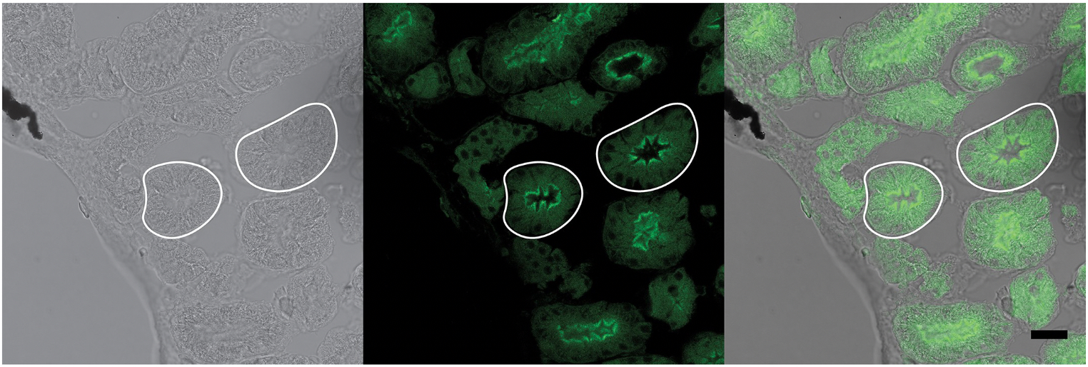
Figure 1. Adult frog kidney slice. Left) DIC image of slice with tubules outlined in white. Middle) Same slice stained with VSP antibody in green, tubules outlined in white. Right) Overlay of DIC and antibody image showing localization of the VSP on the inner (apical) membrane of the tubule.
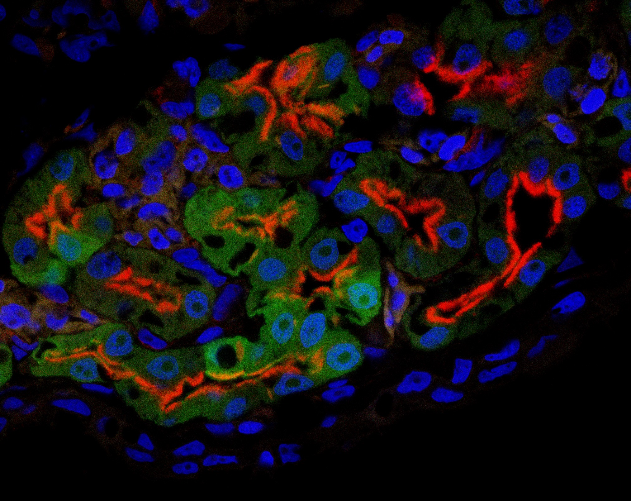
Figure 1. Juvenile frog kidney slice stained with VSP antibody in red and nuclei in blue. Kidney cells are specifically marked with GFP. VSP is found on the apical membrane of the tubules.
We continue to investigate the role of VSP both in the kidney and in the brain!
1. Ratzan W, Rayaprolu V, Killian SE, Bradley R, Kohout SC. The voltage sensing phosphatase (VSP) localizes to the apical membrane of kidney tubule epithelial cells. PLoS One 2019;14:e0209056. https://doi.org/10.1371/journal.pone.0209056. PubMed PMID: 30964862; PubMed Central PMCID: PMC6456211.