Art of Research

Art of Research
Art of Rowan Research Contest & Exhibition
The Art of Rowan Research Contest & Exhibition celebrates student-created imagery revealing the artistry of scientific research and creative inquiry underway at Rowan University.
2026 Winners
First place
BaZakht Melego
"Parasaurolophus: Cloaca & reproductive anatomy"
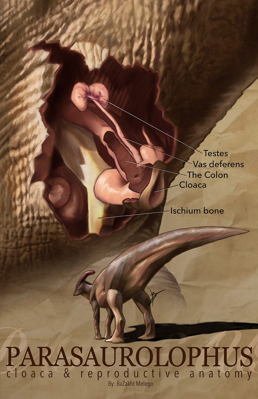 This study proposes an evidence-based reconstruction of the cloacal and reproductive anatomy of a male Parasaurolophus. While soft tissue preservation in non-avian dinosaurs is rare, the documented cloaca of Psittacosaurus confirmed that dinosaurs possessed cloacal structures comparable to modern crocodilians. However, this specimen represents a small ceratopsian lineage, leaving open questions about anatomical variation across larger and more derived taxa such as hadrosaurids.
I initially chose this subject with a degree of humor, fully aware that dinosaur reproductive anatomy occupies an awkward space in both academic and popular discourse. However, the topic is scientifically legitimate and arguably necessary. Given significant differences in body size, pelvic morphology, and inferred behavior, Parasaurolophus may have exhibited distinct cloacal and reproductive adaptations. This project applies phylogenetic bracketing using extant archosaurs (birds and crocodilians), alongside osteology correlates such as pelvic structure, chevron placement, and tail-base musculature, to reconstruct plausible male anatomy.
Inspired by the anatomical clarity of Ute Behrend, the study approaches reproductive biology as an essential component of functional anatomy. By visualizing soft tissue structures grounded in comparative evidence, this work aims to expand palaeobiological understanding beyond skeletal remains and contribute to a more complete reconstruction of dinosaur biology.
Second place
Ryan Victor
"Cicindela columbica"
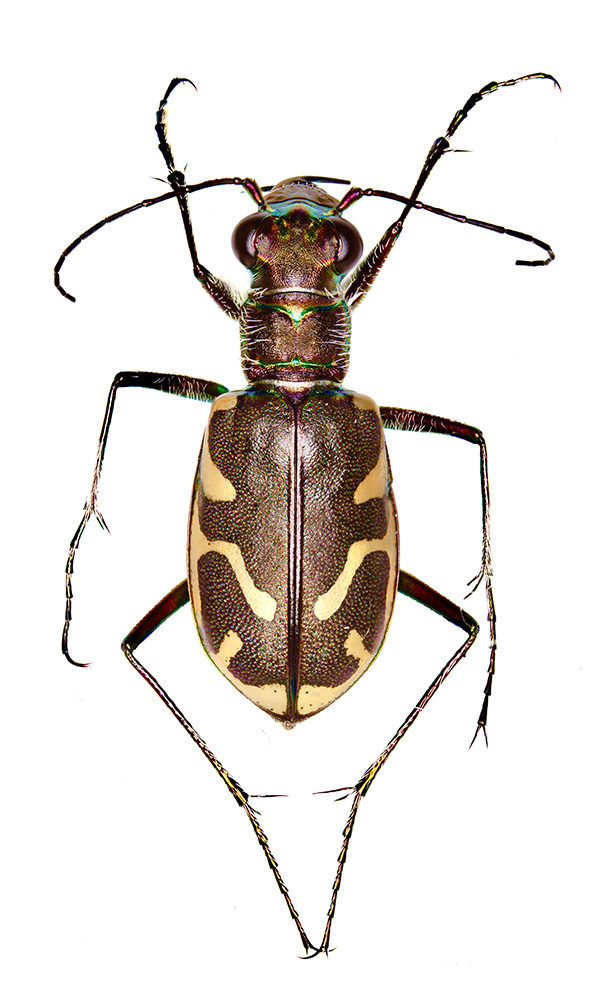
This photo depicts a rare and endangered insect, the Columbia river tiger beetle (Cicindela columbica). Tiger beetles are a remarkable family of insect predators, known for their swift movement and voracious hunting. There are many species of tiger beetles (122 in the US) and some species are difficult to tell apart, sometimes requiring the use of a microscope.
The high resolution of this macro photo allowed us to see key differences in size, shape and the presence of small hairs (setae) in specific places. These subtle differences, which are difficult to see with the naked eye, separate the Columbia river tiger beetle from a common, nearly identical relative, the bronzed tiger beetle (Cicindela repanda). Due to this subtlety, field sightings of this threatened species often go unnoticed.
Most importantly, this photo allowed us to confirm the presence of the Columbia river tiger beetle in an area where it was thought to be locally extinct. Documenting and reporting this sighting provides valuable data which is not commonly available, as well as paving the way for new conservation efforts.
This photo was taken using a macro lens at multiple focal depths, and then processed using focus stacking software, allowing every structure in the image to be in focus simultaneously.
Third place
Angelena Revas
"Billion Year Butterfly"
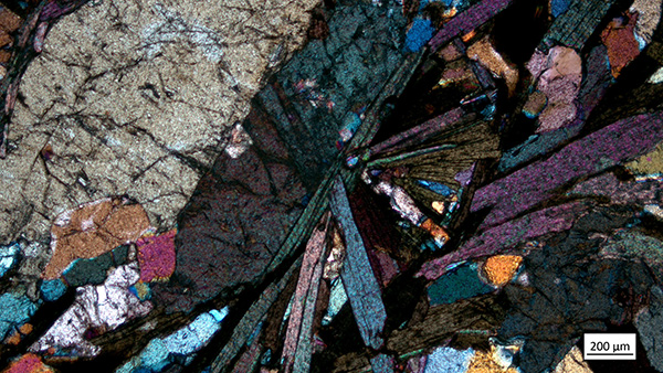
The Appalachian Mountains were created from continental collision between (what is now) North America and Africa during the formation of the last supercontinent, Pangea. The once large mountain chain uplifted older rock to the surface, including Precambrian continental crust around 1billion years old. This collision changed the mineral and textural composition of these rocks: grains aligned, foliation formed, and crystals grew.
During my research with Dr. Lily Pfeifer and Dr. Aaron Barth, I collected samples of said Pennsylvanian bedrock. Geochemistry was analyzed and compared to that of western sediment basins that hold eroded pieces of the ancient Appalachian Mountains in order to constrain weathering trends. To analyze the mineralogy of our samples I developed an in-house method to create petrographic thin sections. Thin sections are around .03 mm thick pieces of rock that are cut to analyze microscopic textures and minerals. Minerals can be identified by key characteristics seen when cross-polarized light shines through the sample like a stained-glass window.
This image shows a microscopic view of the Wissahickon schist, a local geologic formation, under cross-polarized light. Mica crystals display a radial pattern emitting from another vertical crystal creating the image of a butterfly frozen in time.
People's choice
Mohamad Keblawi
"Fluorescent Coalescent"
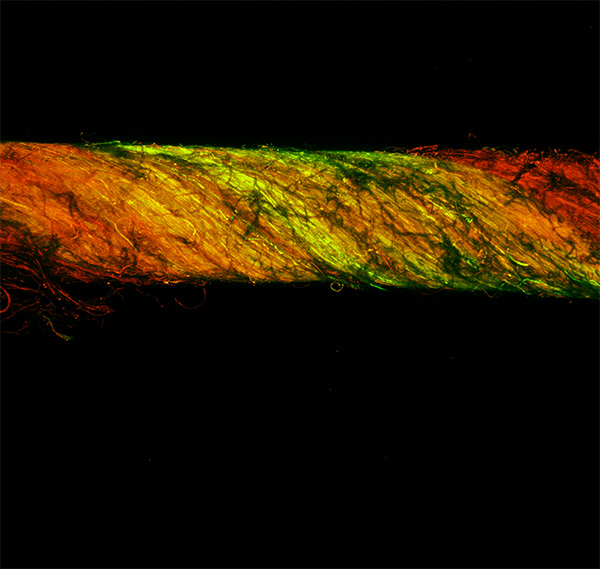
The research established a new method of fabricating yarns using nanofibers as base filaments. The yarn in the image was originally fabricated to demonstrate that multiple different yarn segments can possess an independent set of properties. In this case, one yarn segment fluoresces green while the other fluoresces red. However, the set of properties can be anything from filament density, filament diameter, or twist percentage. The image was captured at a region where the two fluorescent yarn segments intertwine. It was captured using a confocal microscope. The yarn was subjected to a laser beam that caused it to emit a green color, and another laser that caused it to emit a red color. Red and green images where then composited together to produce the image you see right here. The title "Fluorescent Coalescent" is an homage to the Arctic Monkeys song " Fluorescent Adolescent", which happens to be one of my favorite songs.
2026 Student Submissions
This year, Rowan students submitted 42 images for judges to weigh based on their artistic and scientific value. Students, faculty and staff voted to award the People's Choice from among the remaining qualified entries.2026 Contest Timeline
Submissions: Accepted through Feb. 25, 2026
Judging begins: Feb. 26, 2026
Winners notified: Mid-March 2026
Award ceremony and exhibit: Rowan Research Day, Student Center, March 25, 2026
Prizes and recognition
Prizes:
First place: $500
Second place: $300
Third place: $100
People's Choice: Rowan swag bag
Winners were announced during Rowan Research Day on March 25, 2026. Selected images will be featured on Rowan Today, showcased on Rowan University’s website and various publications, and displayed for public view on campus.
Who may submit
Open to all students of the Rowan University community.
What can be submitted
Submissions must be original images of research affiliated with Rowan University. Images may include color-enhanced microscopy, photographs taken in the lab or in the field, graphic illustrations or drawings, fractal imagery or other photos revealing the artistry that is inherent in research, scholarship and creative activity.
Submission rules and guidelines
Photographs or illustrations must be your original work. Any use of artificial intelligence or digital tools to create or enhance the image must be detailed in your description with the submission.
Entries should be a minimum of 5 MB.
Maximum upload size for images is 25 MB.
Photo resolution must be a minimum of 300 dpi and at least 1,500 pixels wide.
Low-resolution images may be disqualified.
Files must be submitted as JPEG or JPG or TIFF.
Images must not contain watermarks.
Images of microbiological material, cells or microorganisms are permitted.
Images must not contain any personally identifiable information.
Images must not be derived from patient data or samples.
Images of laboratory animals are not permitted and will not be judged.
For more information on the rules and guidelines, email us at artofresearch@rowan.edu.
There is no fee to apply.
Submissions must include a short title and a brief (150-300 words) description of the research the image is portraying, written in language non-researchers can understand. Include the name of the project’s principal investigator (PI) and any explanation of what was involved in capturing the photograph or illustration. If applicable, please secure permission for your image from the project’s PI at Rowan, prior to submitting.
Submissions must be relative to current or ongoing projects, with no entries of work created prior to January 2025. Limit is five entries per person.
Images from the same research project may not win more than once.
No person can win more than one prize.
Any operation of an unmanned aerial vehicle (UAV/drone) by a contestant must be in compliance with laws and policies.
Enter the contest
Judging
Judges:
- Tabbetha Dobbins, Ph.D., professor of physics and dean of the School of Graduate Studies
- Lori Marshall, assistant vice president, University Communication
- Mary Salvante, director and chief curator, Rowan University Art Gallery & Museum
- Andrew Hottle, professor of art history and communication studies, Ric Edelman College of Communication, Humanities & Social Sciences
Evaluation criteria
Works will be judged based on their artistic and scientific value.
Where images may be displayed
All entries submitted shall remain the property of the artist who submitted the entry. However, contest submissions may be displayed and used in the following ways with full credit given to the creators:
- Displayed for public exhibit on the Rowan campus
- Posted on Rowan websites
- Posted on Rowan social media accounts
- Reproduced in Rowan’s printed materials including magazines, brochures, signage and promotional materials
Questions about the contest? Email us at artofresearch@rowan.edu.